Myelogram
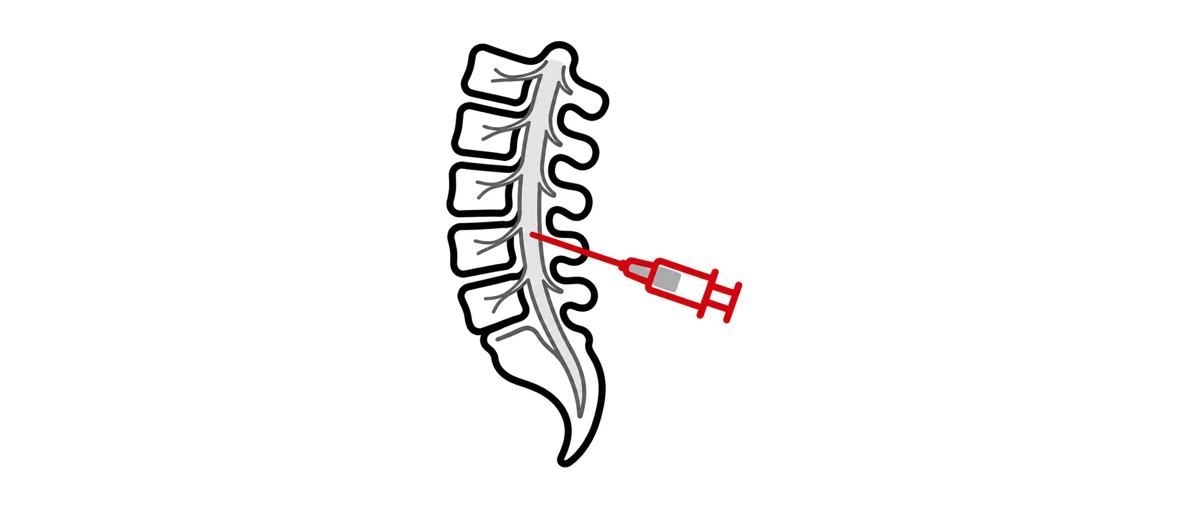
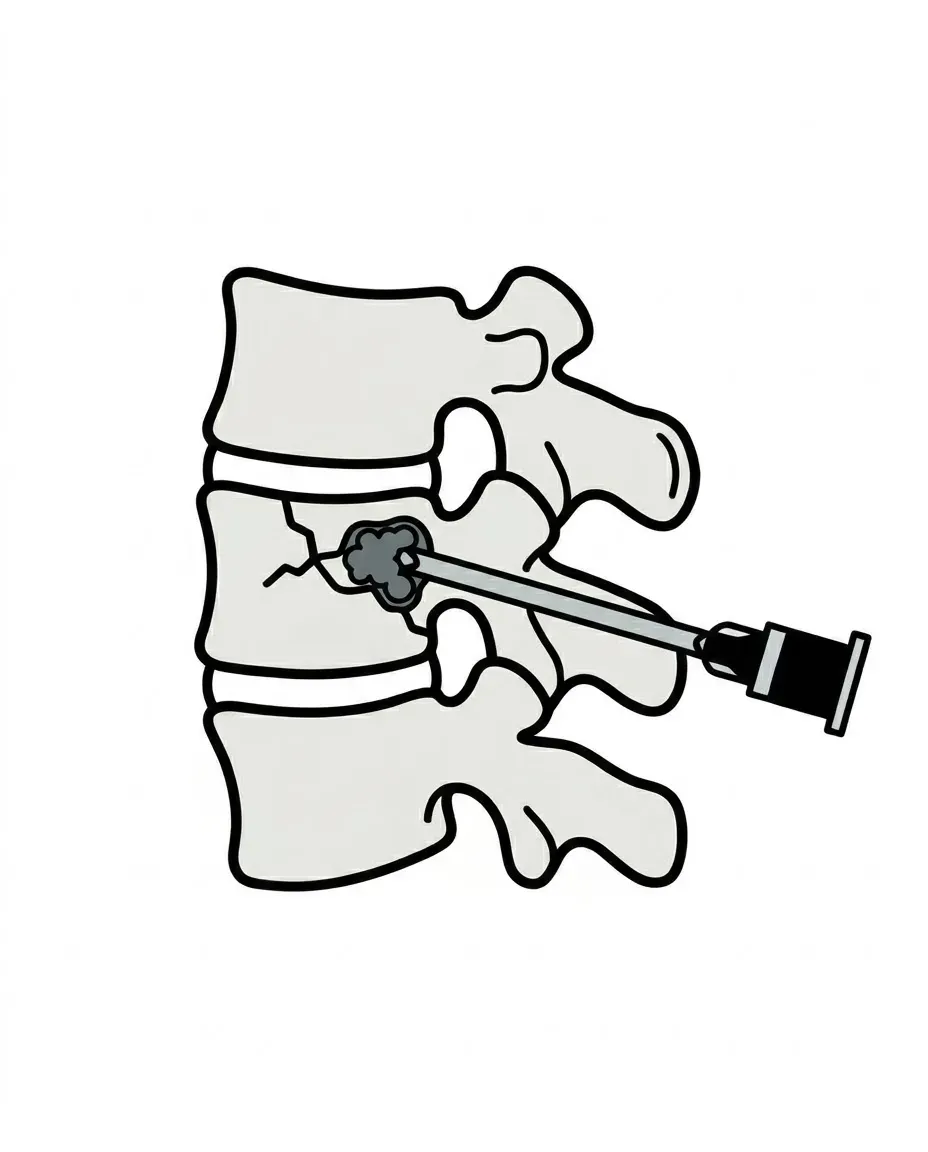
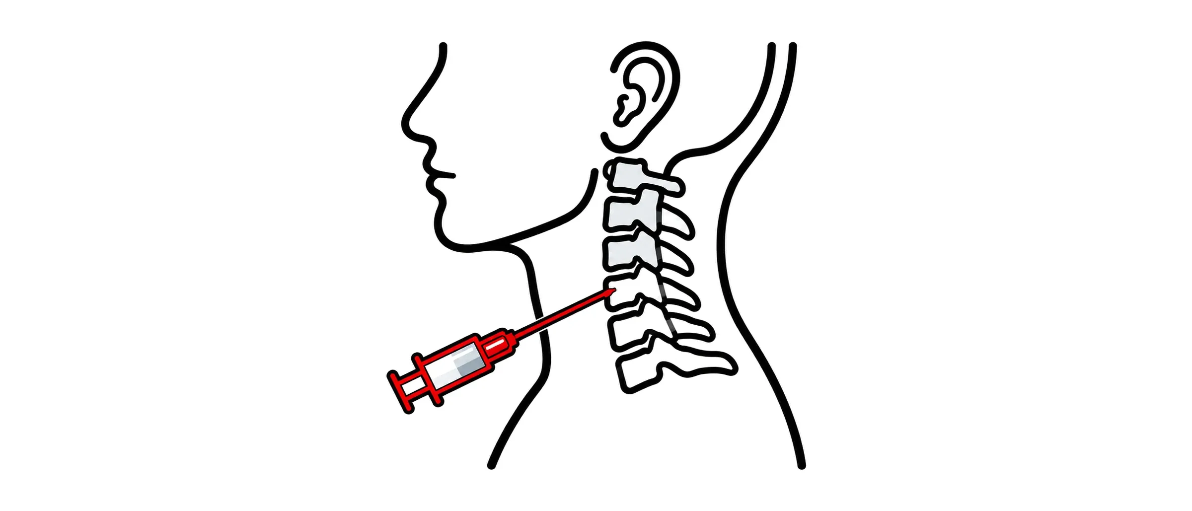
A myelogram is an X-ray or CT study of the spinal canal after contrast dye has been injected into the spinal fluid. It shows the fluid space, the nerve roots and any leak in a way MRI cannot.
Why it is done
I use myelography for two main reasons. The first is to find a spinal CSF leak in spontaneous intracranial hypotension, where dynamic myelography, imaging the contrast as it moves, is the only way to catch a fast leak or a CSF venous fistula. The second is to assess nerve root or cord compression when MRI is not possible (a pacemaker or spinal metalwork, for instance) or is unclear.
What happens
The test is done in the CT or fluoroscopy room, usually as a day case. You lie on your front or side. After local anaesthetic, a fine needle is placed into the spinal fluid in the lower back under imaging guidance, and contrast is injected. For a dynamic study the table is tilted so the contrast runs along the spine while images are taken rapidly; for a standard CT myelogram you are scanned once the contrast has spread. The whole visit takes two to three hours, most of it observation afterwards.
Afterwards
You lie flat for a period, drink plenty and go home with someone. By employing small-gauge, non-cutting needles under direct image guidance, the risk of a post-dural-puncture headache (PDPH) is kept below 5%. If a headache does occur in the following days, it is the commonest after-effect and usually settles with rest and fluids; if it persists, contact us, because it can be treated. Bruising at the needle site is minor. Serious problems, such as infection or a reaction to the contrast, are rare.
Common questions about myelogram
Is it painful?
The local anaesthetic stings briefly. Most people describe pressure rather than pain during the injection.
Why can’t MRI do this?
MRI shows the fluid but not its movement. A leak that empties in seconds, or a fistula into a vein, is invisible unless you watch the contrast flow.
What preparation is needed?
Tell us about blood thinners and any allergy to contrast. Some medicines are paused beforehand; I will tell you which.
What happens if a leak is found?
The myelogram shows exactly where it is, and that decides the treatment. Options include a targeted blood patch or fibrin glue at the leak site, closing a CSF venous fistula from inside the vein or surgical repair. I explain which suits your leak once the images are reviewed.
When will I get the results?
The images are reviewed after the test and the report goes to you and your referring doctor. We then meet to plan the next steps.
Have another question?
Ask a question and the AI assistant will answer from Dr Makalanda’s published information. It cannot give personal medical advice, so please do not share personal or medical details. For emergencies, call 999.