Cranial dural fistula

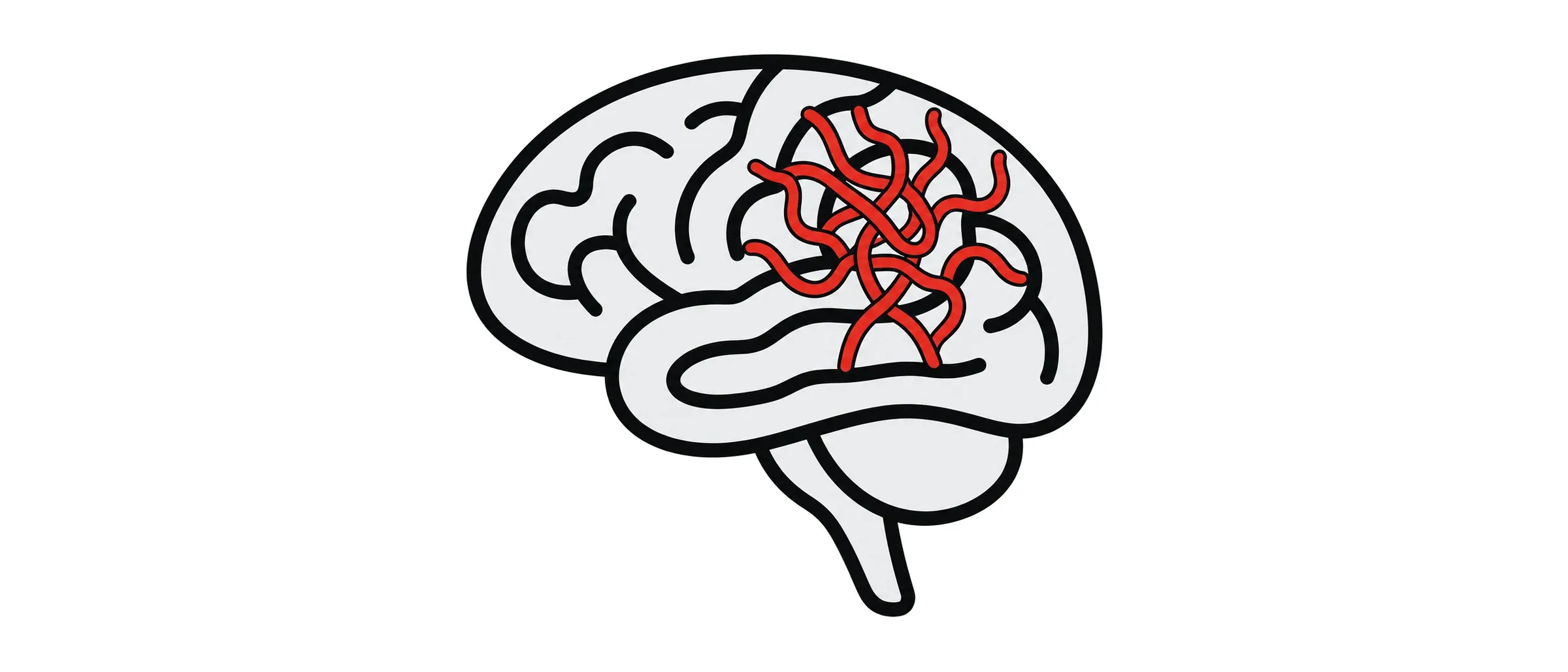
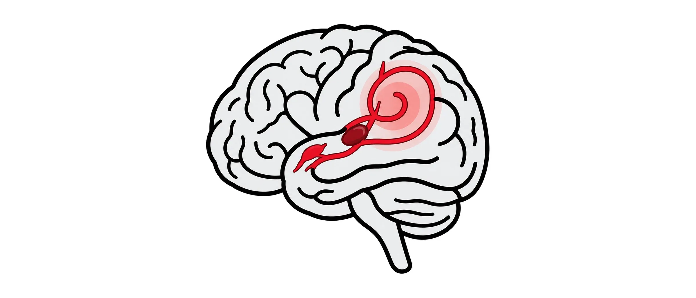
A dural arteriovenous fistula is a direct connection between an artery and a vein in the tough lining around the brain. Some are harmless and only noisy. Some put pressure on the brain’s veins and can cause bleeding. Telling them apart is the point of seeing a specialist.
What it is
Normally blood passes from arteries through tiny capillaries before reaching veins. In a fistula the artery empties straight into a vein, so the vein carries high-pressure arterial blood it was not built for. Fistulas develop later in life, sometimes after a clot in a venous sinus or a head injury, often for no clear reason. The commonest symptom is pulsatile tinnitus, a whooshing in one ear in time with the heartbeat. Fistulas near the eye cause a red, bulging eye or double vision. Fistulas that drain into veins on the surface of the brain can cause seizures, confusion, weakness or a brain haemorrhage, and these are the ones that need treating promptly.
How it is found
MRI and CT angiography raise the suspicion. A catheter angiogram is essential: it shows exactly which arteries feed the fistula and, more importantly, which veins it drains into, because the drainage pattern decides how dangerous it is.
How I treat it
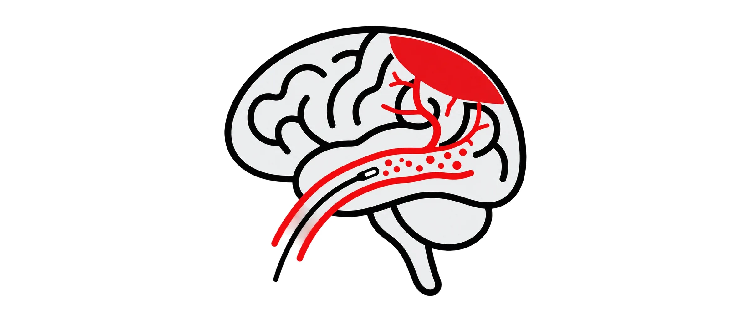
I treat fistulas draining into brain veins, or those causing symptoms you cannot live with. The treatment is embolisation: under general anaesthetic, I navigate a microcatheter from the wrist or groin into the feeding artery or the draining vein and inject a liquid embolic agent or place coils to close the connection. Some fistulas need more than one session. A few are better closed surgically or with focused radiation, and I refer for that when it is the right answer. Low-risk fistulas that are merely noisy can be watched, and I will explain the warning signs to look for.
What to expect
Usually one night in hospital. Most people return to normal activities within a week.
Risks and follow-up
The main risks are stroke, bleeding and damage to the nerves that run near some fistulas, particularly those around the eye and ear. In experienced centres, benchmark figures for procedural risk show that permanent neurological deficit occurs in less than 8% to 10% of cases. I quote the specific figures for your fistula at the consultation. A follow-up angiogram some months later confirms the fistula has stayed closed.
Common questions about cranial dural fistula
The noise is unbearable. Can it be cured?
When the fistula is closed the noise usually stops. Whether treatment is worth the risk for a fistula that is only noisy is a decision we make together.
Can it come back?
Occasionally. That is why I check with follow-up imaging.
Have another question?
Ask a question and the AI assistant will answer from Dr Makalanda’s published information. It cannot give personal medical advice, so please do not share personal or medical details. For emergencies, call 999.