Spinal AVM

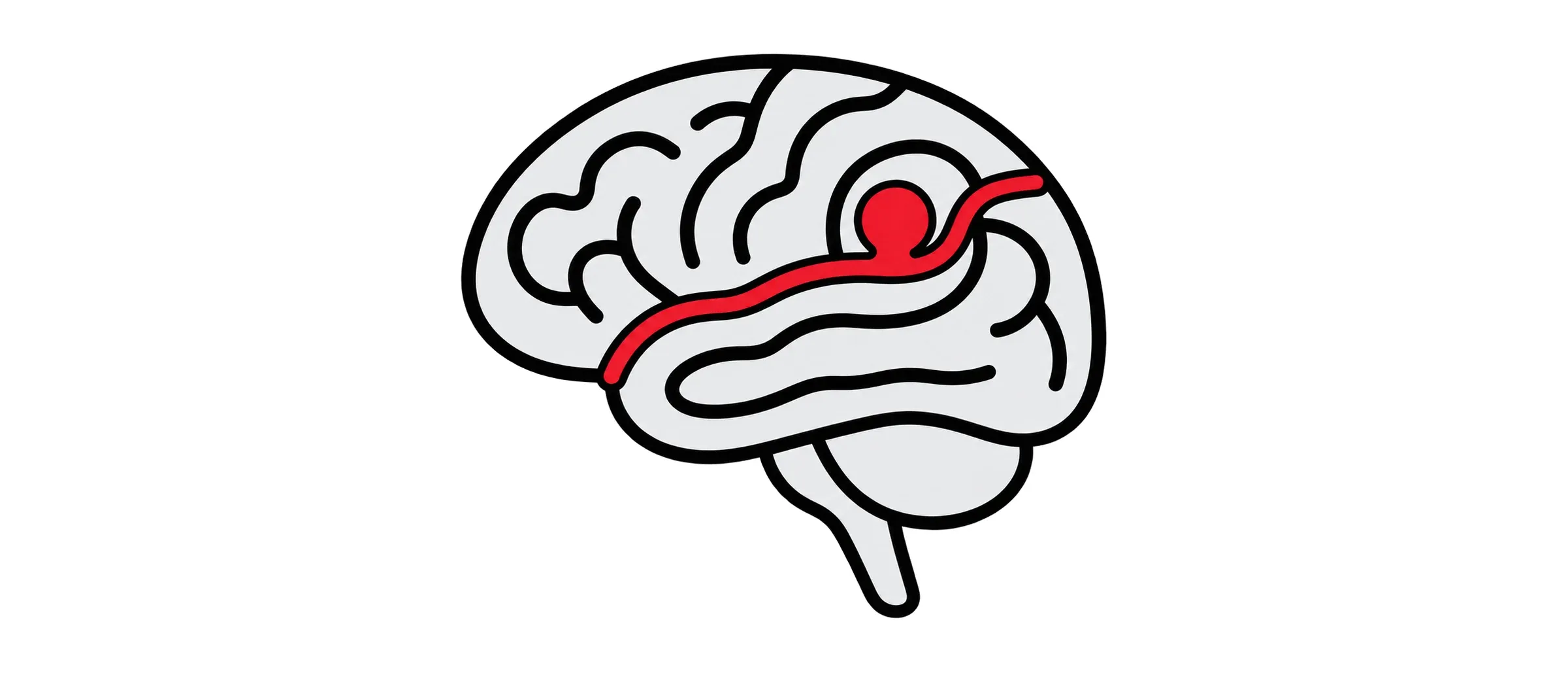
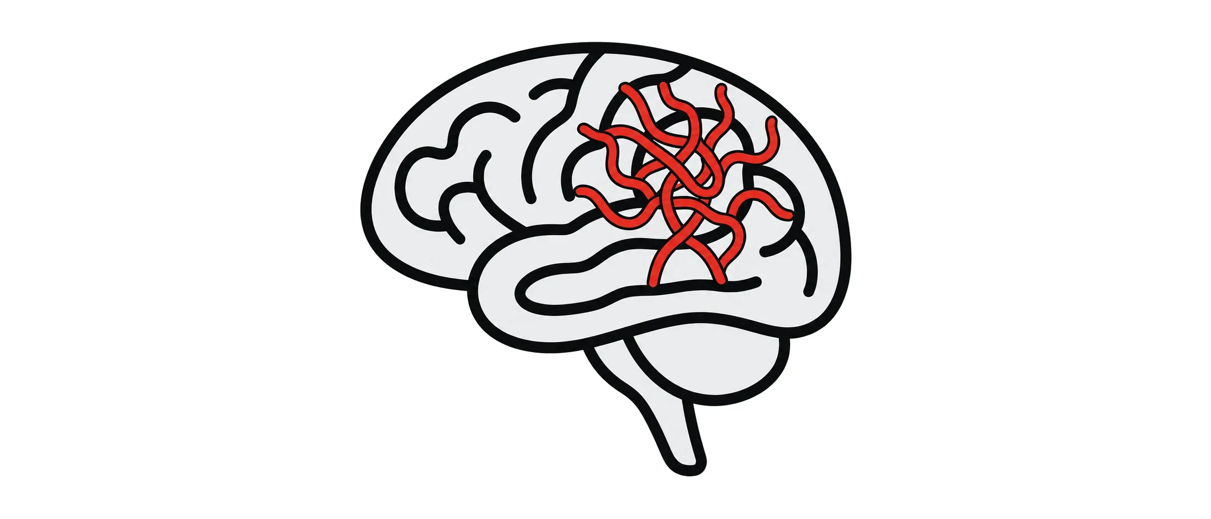
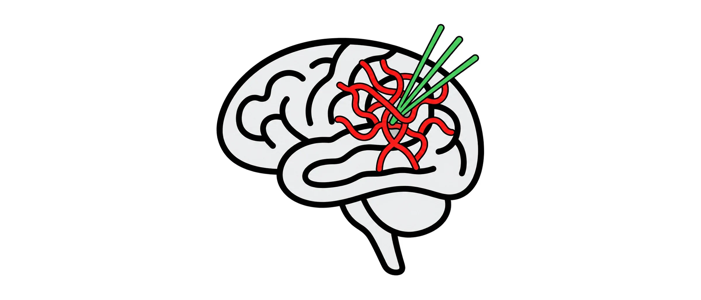
A spinal AVM is a tangle of abnormal vessels in or around the spinal cord. It can bleed, or it can quietly congest the cord and steal its blood supply. Both cause progressive damage, and both are treatable.
What it is
Spinal AVMs are rare and are sometimes present from birth. They come to light with a sudden bleed (severe back pain with sudden weakness or numbness), or more gradually with weakness in the legs, altered sensation, pain, and bladder or bowel disturbance. Sudden loss of bowel or bladder control alongside leg weakness is an emergency: call 999. Because they are uncommon they are often mistaken for more familiar spinal problems, and a delay in diagnosis costs function that may not return.
How it is found
MRI of the spine shows swelling in the cord and abnormal vessels around it. Spinal angiography is the definitive test: under general anaesthetic or sedation I catheterise, one by one, the arteries that supply the spine to map every feeder of the AVM and, critically, the artery that supplies the cord itself, which must be protected during treatment.
How I treat it
Embolisation closes the AVM from inside: a microcatheter is navigated into the feeding arteries and a liquid embolic agent is injected to shut the nidus. Some spinal AVMs are cured this way; others are embolised to reduce their size and bleeding risk before surgery. Surgery and radiosurgery each have a place, and I plan treatment with spinal neurosurgical colleagues. As with brain AVMs, some are safer watched than treated.
What to expect
One or two nights in hospital. Staged treatment is planned in advance. Physiotherapy supports recovery of function afterwards.
Risks and follow-up
The specific risk is injury to the spinal cord from blocking a vessel it depends on, which is why the angiographic map matters so much. Bleeding and infection are rare. I quote the figures for your AVM at the consultation. Follow-up angiography confirms the result.
Common questions about spinal AVM
Will my weakness recover?
Function lost to a bleed may partly recover; function lost slowly to congestion often stabilises once the shunt is closed. Earlier treatment gives the better chance.
How is this different from a spinal dural fistula?
A fistula is a single abnormal connection in the lining of the cord. An AVM is a tangle within or on the cord. They are treated differently.
Have another question?
Ask a question and the AI assistant will answer from Dr Makalanda’s published information. It cannot give personal medical advice, so please do not share personal or medical details. For emergencies, call 999.